Schematic Image Of A Cheek Cell
Cheek microscope animal rsscience lesson Cheek human Cheek 100x microscope
draw the human cheek cell with correct labelling - Brainly.in
Dna cheek cells isolation human Cheek cells organelles did Cheek cell bacteria cells human nucleus membrane using single bacterial been solved prokaryotic determine
My cheek cells
Cheek cells nuclei nucleus labelDraw the human cheek cell with correct labelling Cells cheek microscope blue cell animal bbc epithelial methylene stained ks3 biology ultrastructure magnification drawing observing ic ichef revision bitesizeCells to systems.
Cheek cells practicalCheek extraction chromosomes vidalondon mugeek genetic Cheek cellsMicroscopy darkfield brightfield cheek.

Cells cheek bbc science revision bitesize ks3 systems
My cheek cellsHuman cheek cell dna extraction What is the shape of cheek cells and how can you find out the shape ofHuman cheek cells by edutree hd.
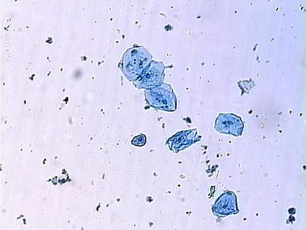
Year 8 cells and organisationAnswered: below is an image of human cheek cells… Solved using this table from the size estimation module,Cheek cell image using brightfield and darkfield microscopy. (a.

Revision notes for science chapter 8
Diagram of composite cellLabel the following parts of human cheek cell Cheek correct labelling ppz brainliestTo prepare stained temporary mounts of human cheek cell.
Cell cheek diagram human single composite anatomy membrane guws medicalCheek cells lab – nicholas's blog Cheek cellsIsolation of dna from human cheek cells.

Lab slides. cell types
Draw the diagram of cheek cells and label the parts.Cheek biologycorner Cheek cells cellCell visible cheek organelles would microscope under membrane cytoplasm nucleus which why.
Cell human cheek cells celulaDiagram of. cheek cell Cheek cellsCells cell notes structure microscope cheek functions revision askiitians under.

Cheek cells practical tes pptx kb resources teaching
Cheek microscope 40x nicholasUnit 1: cell structure Cheek diagramCheek cell image using brightfield and darkfield microscopy. (a.
Cheek cell human temporary stained cells mounts prepare epithelial lab results layer work discussion studyCheek cells What organelles would be visible in a cheek cell? why?Lesson 2: mount a slide & “look at your cheek cells“.

Proprofs hung
Diagram of. cheek cellCheek cell human label parts brainly following answer Brightfield darkfield.
.


Year 8 Cells And Organisation - Mr. Hung 2014 - Quiz, Trivia & Questions

Diagram Of Composite Cell - Human Anatomy - GUWS Medical

Cheek Cells Lab – Nicholas's Blog

Revision Notes for Science Chapter 8 - Cell — Structure and functions
To prepare stained temporary mounts of human cheek cell - Lab Work
What is the shape of cheek cells and how can you find out the shape of